Home
/ Labeled Diagram Of An - A Diagram Of The Morphology Of A Bird Labeling Different Parts This Download Scientific Diagram _ A fish bone diagram is a common tool used for a cause and effect analysis, where you try to identify possible causes for a certain problem or event.
Labeled Diagram Of An - A Diagram Of The Morphology Of A Bird Labeling Different Parts This Download Scientific Diagram _ A fish bone diagram is a common tool used for a cause and effect analysis, where you try to identify possible causes for a certain problem or event.
Labeled Diagram Of An - A Diagram Of The Morphology Of A Bird Labeling Different Parts This Download Scientific Diagram _ A fish bone diagram is a common tool used for a cause and effect analysis, where you try to identify possible causes for a certain problem or event.. May 31, 2021 · labeled brain diagram. There are three structural parts of the microscope i.e. In this diagram i simply labeled the partitions with the text applicant, registrar, and system although it is also common to put actor symbols (stick figures) to make it very clear that an actor is performing some activities. _____ use the amino acid chart on the last page to identify the amino acids from the mrna sequence in problem # 10. Try to memorize the name and location of each structure, then proceed to test yourself with the blank brain diagram provided below.
10) the sense strand of a dna molecule is: A fish bone diagram is a common tool used for a cause and effect analysis, where you try to identify possible causes for a certain problem or event. Before exploring microscope parts and functions, you should probably understand that the compound light microscope is more complicated than just a microscope with more than one lens. A fishbone diagram is another name for the ishikawa diagram or cause and effect diagram.it gets its name from the fact that the shape looks a bit like a fish skeleton. Jul 01, 2021 · figure created with biorender.com.
A Labeled Diagram Of The Pancreas Stock Illustration Download Image Now Istock from media.istockphoto.com Jul 01, 2021 · figure created with biorender.com. Diagram of parts of a microscope. May 31, 2021 · labeled brain diagram. Microscope parts and functions with labeled diagram and functions how does a compound microscope work?. Ystems thinking has been described as a language for talking about the complex, interdependent issues managers face every day. Try to memorize the name and location of each structure, then proceed to test yourself with the blank brain diagram provided below. First up, have a look at the labeled brain structures on the image below. Identify the labeled structures on the following diagram of translation.
A labeled tree has specific values assigned to its leaves, while an unlabeled tree, sometimes called a tree shape, defines a topology only.
On the pixel 3 xl, the cameras are on either side of the top speaker. C c c a c g t c t the mrna sequence from this dna molecule is : Microscope parts and functions with labeled diagram and functions how does a compound microscope work?. On the pixel 3, both cameras are on the top left. Jul 01, 2021 · figure created with biorender.com. In this diagram i simply labeled the partitions with the text applicant, registrar, and system although it is also common to put actor symbols (stick figures) to make it very clear that an actor is performing some activities. Ystems thinking has been described as a language for talking about the complex, interdependent issues managers face every day. Try to memorize the name and location of each structure, then proceed to test yourself with the blank brain diagram provided below. 10) the sense strand of a dna molecule is: May 31, 2021 · labeled brain diagram. First up, have a look at the labeled brain structures on the image below. Diagram of parts of a microscope. Before exploring microscope parts and functions, you should probably understand that the compound light microscope is more complicated than just a microscope with more than one lens.
On the pixel 3 xl, the cameras are on either side of the top speaker. _____ use the amino acid chart on the last page to identify the amino acids from the mrna sequence in problem # 10. Diagram of parts of a microscope. Before exploring microscope parts and functions, you should probably understand that the compound light microscope is more complicated than just a microscope with more than one lens. Try to memorize the name and location of each structure, then proceed to test yourself with the blank brain diagram provided below.
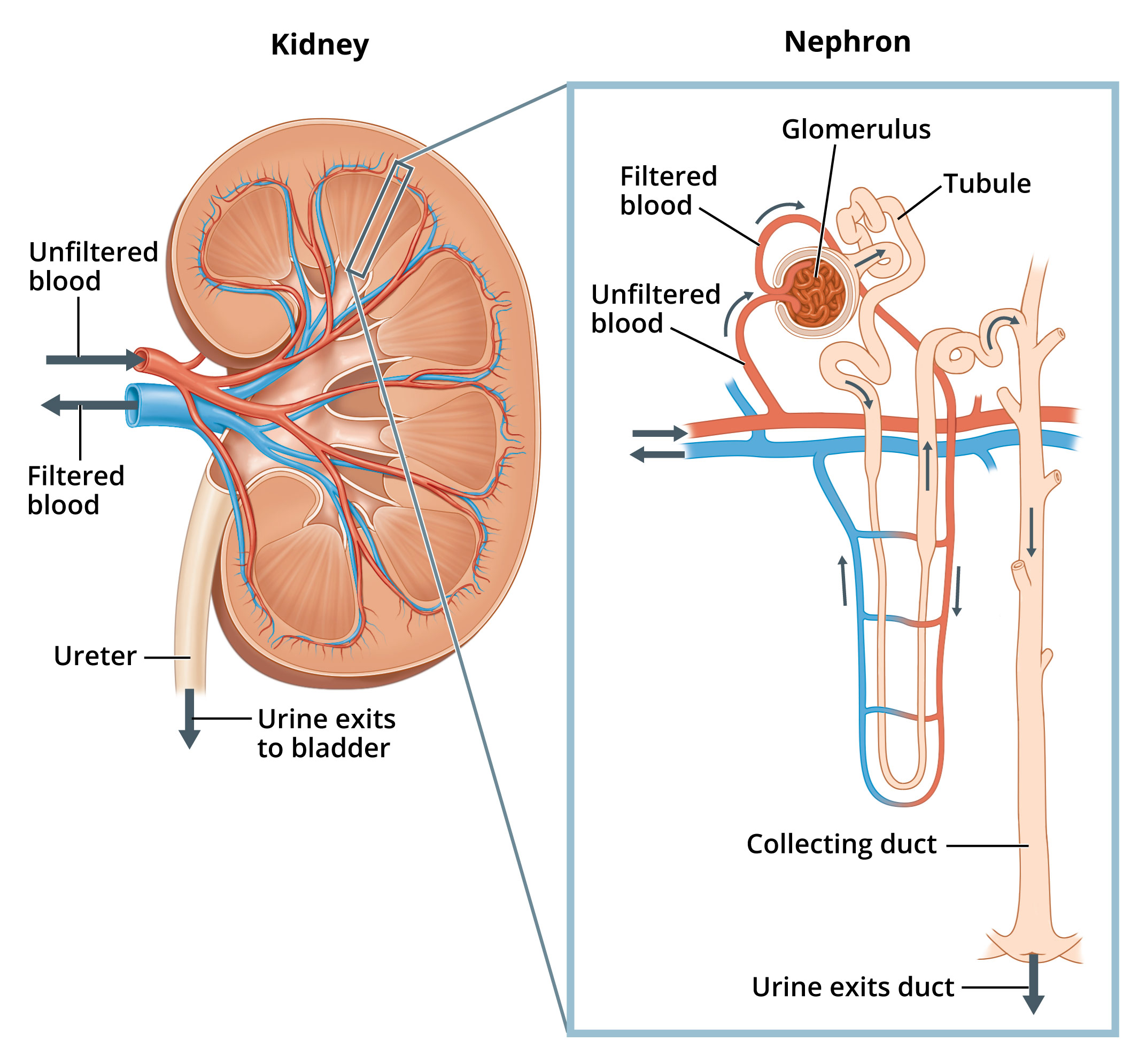
Nephron Labeled Diagram Anatomy And Structure from sciencediagrams.com May 31, 2021 · labeled brain diagram. A labeled tree has specific values assigned to its leaves, while an unlabeled tree, sometimes called a tree shape, defines a topology only. C c c a c g t c t the mrna sequence from this dna molecule is : Before exploring microscope parts and functions, you should probably understand that the compound light microscope is more complicated than just a microscope with more than one lens. There are three structural parts of the microscope i.e. First up, have a look at the labeled brain structures on the image below. Microscope parts and functions with labeled diagram and functions how does a compound microscope work?. A fish bone diagram is a common tool used for a cause and effect analysis, where you try to identify possible causes for a certain problem or event.
Before exploring microscope parts and functions, you should probably understand that the compound light microscope is more complicated than just a microscope with more than one lens.
There are three structural parts of the microscope i.e. _____ use the amino acid chart on the last page to identify the amino acids from the mrna sequence in problem # 10. Microscope parts and functions with labeled diagram and functions how does a compound microscope work?. Diagram of parts of a microscope. A fishbone diagram is another name for the ishikawa diagram or cause and effect diagram.it gets its name from the fact that the shape looks a bit like a fish skeleton. Identify the labeled structures on the following diagram of translation. C c c a c g t c t the mrna sequence from this dna molecule is : 10) the sense strand of a dna molecule is: On the pixel 3, both cameras are on the top left. Within that framework, causal loop diagrams can be thought of as sentences that are constructed by identifying the key variables in a system (the "nouns") and indicating the causal relationships between them via links (the "verbs"). Ystems thinking has been described as a language for talking about the complex, interdependent issues managers face every day. Jul 01, 2021 · figure created with biorender.com. Before exploring microscope parts and functions, you should probably understand that the compound light microscope is more complicated than just a microscope with more than one lens.
A fishbone diagram is another name for the ishikawa diagram or cause and effect diagram.it gets its name from the fact that the shape looks a bit like a fish skeleton. A fish bone diagram is a common tool used for a cause and effect analysis, where you try to identify possible causes for a certain problem or event. In this diagram i simply labeled the partitions with the text applicant, registrar, and system although it is also common to put actor symbols (stick figures) to make it very clear that an actor is performing some activities. First up, have a look at the labeled brain structures on the image below. Identify the labeled structures on the following diagram of translation.
Labeled Diagram Of The Cardiovascular System Download Scientific Diagram from www.researchgate.net A labeled tree has specific values assigned to its leaves, while an unlabeled tree, sometimes called a tree shape, defines a topology only. On the pixel 3 xl, the cameras are on either side of the top speaker. _____ use the amino acid chart on the last page to identify the amino acids from the mrna sequence in problem # 10. There are three structural parts of the microscope i.e. Microscope parts and functions with labeled diagram and functions how does a compound microscope work?. Diagram of parts of a microscope. Try to memorize the name and location of each structure, then proceed to test yourself with the blank brain diagram provided below. On the pixel 3, both cameras are on the top left.
First up, have a look at the labeled brain structures on the image below.
Identify the labeled structures on the following diagram of translation. Try to memorize the name and location of each structure, then proceed to test yourself with the blank brain diagram provided below. There are three structural parts of the microscope i.e. C c c a c g t c t the mrna sequence from this dna molecule is : Within that framework, causal loop diagrams can be thought of as sentences that are constructed by identifying the key variables in a system (the "nouns") and indicating the causal relationships between them via links (the "verbs"). Diagram of parts of a microscope. Before exploring microscope parts and functions, you should probably understand that the compound light microscope is more complicated than just a microscope with more than one lens. Microscope parts and functions with labeled diagram and functions how does a compound microscope work?. On the pixel 3 xl, the cameras are on either side of the top speaker. In this diagram i simply labeled the partitions with the text applicant, registrar, and system although it is also common to put actor symbols (stick figures) to make it very clear that an actor is performing some activities. Ystems thinking has been described as a language for talking about the complex, interdependent issues managers face every day. 10) the sense strand of a dna molecule is: A fishbone diagram is another name for the ishikawa diagram or cause and effect diagram.it gets its name from the fact that the shape looks a bit like a fish skeleton.